| Weight | 1 lbs |
|---|---|
| Dimensions | 9 × 5 × 2 in |
| accession | P54764 |
| express system | HEK293 |
| product tag | C-His |
| purity | > 95% as determined by Tris-Bis PAGE;> 95% as determined by HPLC |
| background | The expression and activation of EphA4 in the various cell types in a knee joint was upregulated upon an intraarticular injury. To determine if EphA4 signaling plays a role in osteoarthritis, we determined whether deficient EphA4 expression (in EphA4 knockout mice) or upregulation of the EphA4 signaling (with the EfnA4-fc treatment) would alter cellular functions of synoviocytes and articular chondrocytes. In synoviocytes, deficient EphA4 expression enhanced, whereas activation of the EphA4 signaling reduced, expression and secretion of key inflammatory cytokines and matrix metalloproteases. |
| molecular weight | The protein has a predicted MW of 59.5 kDa. Due to glycosylation, the protein migrates to 60-70 kDa based on Tris-Bis PAGE result. |
| available size | 100 µg, 500 µg |
| endotoxin | Less than 1EU per μg by the LAL method. |

Human EPHA4 Protein 3776
$270.00 – $900.00
Summary
- Expression: HEK293
- Pure: Yes (HPLC)
- Amino Acid Range: Val20-Thr547
Human EPHA4 Protein 3776
| protein |
|---|
| Size and concentration 100, 500µg and lyophilized |
| Form Lyophilized |
| Storage Instructions Valid for 12 months from date of receipt when stored at -80°C. Recommend to aliquot the protein into smaller quantities for optimal storage. Please minimize freeze-thaw cycles. |
| Storage buffer Shipped at ambient temperature. |
| Purity > 95% as determined by Tris-Bis PAGE |
| target relevance |
|---|
| The expression and activation of EphA4 in the various cell types in a knee joint was upregulated upon an intraarticular injury. To determine if EphA4 signaling plays a role in osteoarthritis, we determined whether deficient EphA4 expression (in EphA4 knockout mice) or upregulation of the EphA4 signaling (with the EfnA4-fc treatment) would alter cellular functions of synoviocytes and articular chondrocytes. In synoviocytes, deficient EphA4 expression enhanced, whereas activation of the EphA4 signaling reduced, expression and secretion of key inflammatory cytokines and matrix metalloproteases. |
| Protein names Ephrin type-A receptor 4 (EC 2.7.10.1) (EPH-like kinase 8) (EK8) (hEK8) (Tyrosine-protein kinase TYRO1) (Tyrosine-protein kinase receptor SEK) |
| Gene names EPHA4,EPHA4 HEK8 SEK TYRO1 |
| Protein family Protein kinase superfamily, Tyr protein kinase family, Ephrin receptor subfamily |
| Mass 9606Da |
| Function Receptor tyrosine kinase which binds membrane-bound ephrin family ligands residing on adjacent cells, leading to contact-dependent bidirectional signaling into neighboring cells. The signaling pathway downstream of the receptor is referred to as forward signaling while the signaling pathway downstream of the ephrin ligand is referred to as reverse signaling. Highly promiscuous, it has the unique property among Eph receptors to bind and to be physiologically activated by both GPI-anchored ephrin-A and transmembrane ephrin-B ligands including EFNA1 and EFNB3. Upon activation by ephrin ligands, modulates cell morphology and integrin-dependent cell adhesion through regulation of the Rac, Rap and Rho GTPases activity. Plays an important role in the development of the nervous system controlling different steps of axonal guidance including the establishment of the corticospinal projections. May also control the segregation of motor and sensory axons during neuromuscular circuit development. In addition to its role in axonal guidance plays a role in synaptic plasticity. Activated by EFNA1 phosphorylates CDK5 at 'Tyr-15' which in turn phosphorylates NGEF regulating RHOA and dendritic spine morphogenesis. In the nervous system, also plays a role in repair after injury preventing axonal regeneration and in angiogenesis playing a role in central nervous system vascular formation. Additionally, its promiscuity makes it available to participate in a variety of cell-cell signaling regulating for instance the development of the thymic epithelium. During development of the cochlear organ of Corti, regulates pillar cell separation by forming a ternary complex with ADAM10 and CADH1 which facilitates the cleavage of CADH1 by ADAM10 and disruption of adherens junctions (By similarity). Phosphorylates CAPRIN1, promoting CAPRIN1-dependent formation of a membraneless compartment (By similarity). |
| Catalytic activity #N/A |
| Subellular location Cell membrane ; Single-pass type I membrane protein. Cell projection, axon. Cell projection, dendrite. Postsynaptic density membrane. Early endosome. Cell junction, adherens junction. Note=Clustered upon activation and targeted to early endosome. |
| Tissues Ubiquitous. |
| Structure Heterotetramer upon binding of the ligand. The heterotetramer is composed of an ephrin dimer and a receptor dimer. Oligomerization is probably required to induce biological responses. Interacts (phosphorylated at position Tyr-602) with FYN. Interacts with CDK5, CDK5R1 and NGEF; upon activation by EFNA1 induces NGEF phosphorylation by the kinase CDK5. Interacts with CHN1; effector of EPHA4 in axon guidance linking EPHA4 activation to RAC1 regulation (By similarity). Interacts (via PDZ motif) with SIPA1L1 (via PDZ domain); controls neuronal morphology through regulation of the RAP1 (RAP1A or RAP1B) and RAP2 (RAP2A, RAP2B or RAP2C) GTPases. Forms a ternary complex composed of ADAM10, CADH1 and EPHA4; within the complex, CADH1 is cleaved by ADAM10 which disrupts adherens junctions (By similarity). |
| Domain Th |
| Target Relevance information above includes information from UniProt accession: P54764 |
| The UniProt Consortium |
Data
|
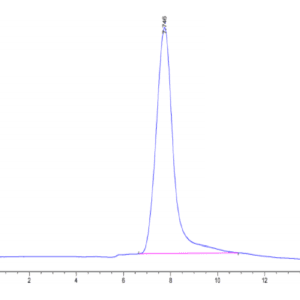
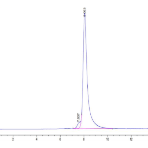
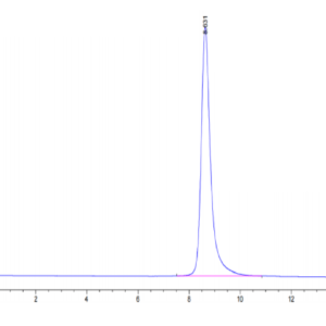
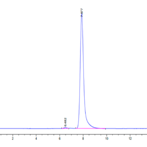
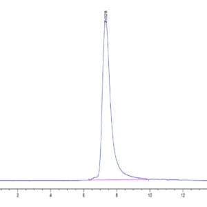
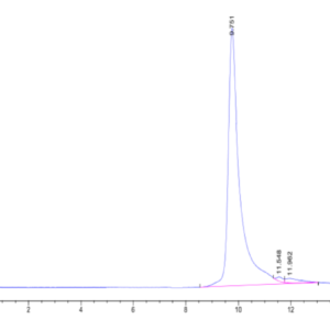
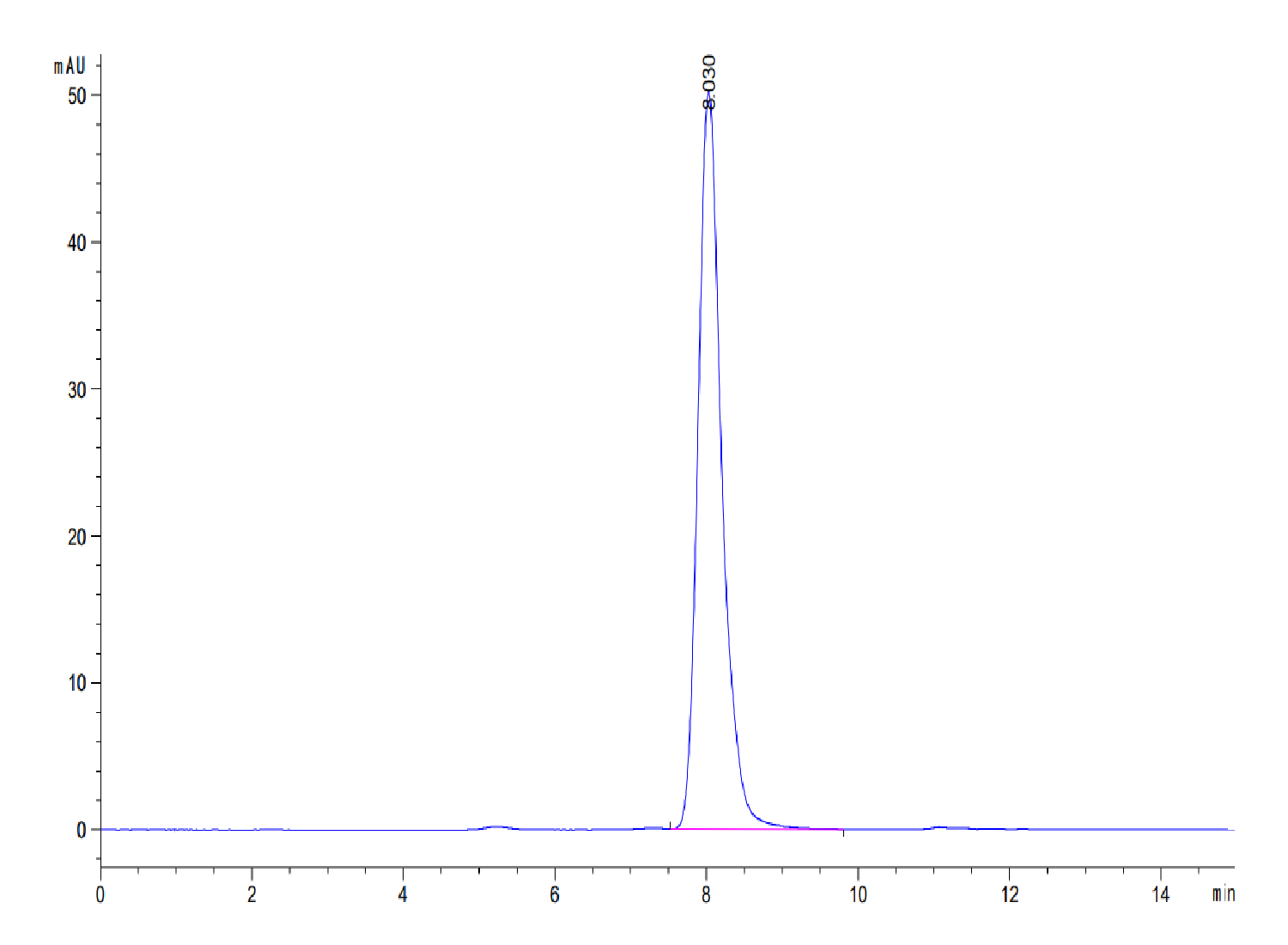
| The purity of Human EPHA4 is greater than 95% as determined by SEC-HPLC. |
|
| Human EPHA4 on Tris-Bis PAGE under reduced condition. The purity is greater than 95%. |
Publications
Publications
| pmid | title | authors | citation |
|---|---|---|---|
| We haven't added any publications to our database yet. | |||
Protocols
| relevant to this product |
|---|
Documents
| # | ||
|---|---|---|
| Please enter your product and batch number here to retrieve product datasheet, SDS, and QC information. | ||