| Weight | 1 lbs |
|---|---|
| Dimensions | 9 × 5 × 2 in |
| accession | O75509 |
| express system | HEK293 |
| product tag | C-His |
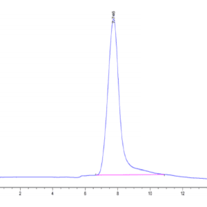
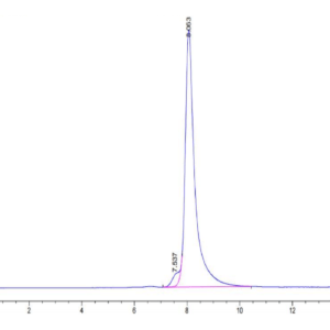
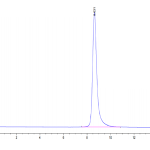
| purity | > 95% as determined by Tris-Bis PAGE |
| background | beta-amyloid precursor protein (APP) and death receptor 6 (DR6, also known as TNFRSF21) activate a widespread caspase-dependent self-destruction program. DR6 is broadly expressed by developing neurons, and is required for normal cell body death and axonal pruning both in vivo and after trophic-factor deprivation in vitro.DR6 is activated locally by an inactive surface ligand(s) that is released in an active form after trophic-factor deprivation. |
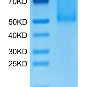
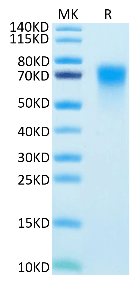
| molecular weight | The protein has a predicted MW of 34.3 kDa. Due to glycosylation, the protein migrates to 65-80 kDa based on Tris-Bis PAGE result. |
| available size | 100 µg, 500 µg |
| endotoxin | Less than 1EU per μg by the LAL method. |
Human DR6/TNFRSF21 Protein 3943
$150.00 – $500.00
Summary
- Expression: HEK293
- Functional: Yes (ELISA)
- Amino Acid Range: Gln42-His349
Human DR6/TNFRSF21 Protein 3943
| protein |
|---|
| Size and concentration 100, 500µg and lyophilized |
| Form Lyophilized |
| Storage Instructions Valid for 12 months from date of receipt when stored at -80°C. Recommend to aliquot the protein into smaller quantities for optimal storage. Please minimize freeze-thaw cycles. |
| Storage buffer Shipped at ambient temperature. |
| Purity > 95% as determined by Tris-Bis PAGE |
| target relevance |
|---|
| beta-amyloid precursor protein (APP) and death receptor 6 (DR6, also known as TNFRSF21) activate a widespread caspase-dependent self-destruction program. DR6 is broadly expressed by developing neurons, and is required for normal cell body death and axonal pruning both in vivo and after trophic-factor deprivation in vitro.DR6 is activated locally by an inactive surface ligand(s) that is released in an active form after trophic-factor deprivation. |
| Protein names Tumor necrosis factor receptor superfamily member 21 (Death receptor 6) (CD antigen CD358) |
| Gene names TNFRSF21,TNFRSF21 DR6 UNQ437/PRO868 |
| Mass 71845Da |
| Function Promotes apoptosis, possibly via a pathway that involves the activation of NF-kappa-B. Can also promote apoptosis mediated by BAX and by the release of cytochrome c from the mitochondria into the cytoplasm. Plays a role in neuronal apoptosis, including apoptosis in response to amyloid peptides derived from APP, and is required for both normal cell body death and axonal pruning. Trophic-factor deprivation triggers the cleavage of surface APP by beta-secretase to release sAPP-beta which is further cleaved to release an N-terminal fragment of APP (N-APP). N-APP binds TNFRSF21; this triggers caspase activation and degeneration of both neuronal cell bodies (via caspase-3) and axons (via caspase-6). Negatively regulates oligodendrocyte survival, maturation and myelination. Plays a role in signaling cascades triggered by stimulation of T-cell receptors, in the adaptive immune response and in the regulation of T-cell differentiation and proliferation. Negatively regulates T-cell responses and the release of cytokines such as IL4, IL5, IL10, IL13 and IFNG by Th2 cells. Negatively regulates the production of IgG, IgM and IgM in response to antigens. May inhibit the activation of JNK in response to T-cell stimulation. Also acts as a regulator of pyroptosis: recruits CASP8 in response to reactive oxygen species (ROS) and subsequent oxidation, leading to activation of GSDMC (PubMed:34012073). |
| Subellular location Cell membrane ; Single-pass type I membrane protein. Note=Endocytosed following oxidation in response to reactive oxygen species (ROS). |
| Tissues Detected in fetal spinal cord and in brain neurons, with higher levels in brain from Alzheimer disease patients (at protein level). Highly expressed in heart, brain, placenta, pancreas, lymph node, thymus and prostate. Detected at lower levels in lung, skeletal muscle, kidney, testis, uterus, small intestine, colon, spleen, bone marrow and fetal liver. Very low levels were found in adult liver and peripheral blood leukocytes. |
| Structure Associates with TRADD (PubMed:9714541). Interacts with NGFR (PubMed:23559013). Interacts with CASP8 (PubMed:34012073). Interacts with N-APP (By similarity).; (Microbial infection) Interacts with hepatitis C virus (HCV) non-structural protein 5A; this interaction allows the modulation by the virus of JNK, p38 MAPK, STAT3, and Akt signaling pathways in a DR6-dependent manner. |
| Post-translational modification Oxidized in response to reactive oxygen species (ROS), leading to endocytosis. |
| Domain TOPO_DOM 4 |
| Target Relevance information above includes information from UniProt accession: O75509 |
| The UniProt Consortium |
Data
|
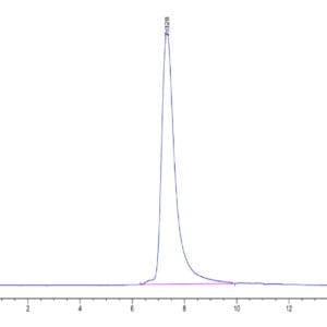
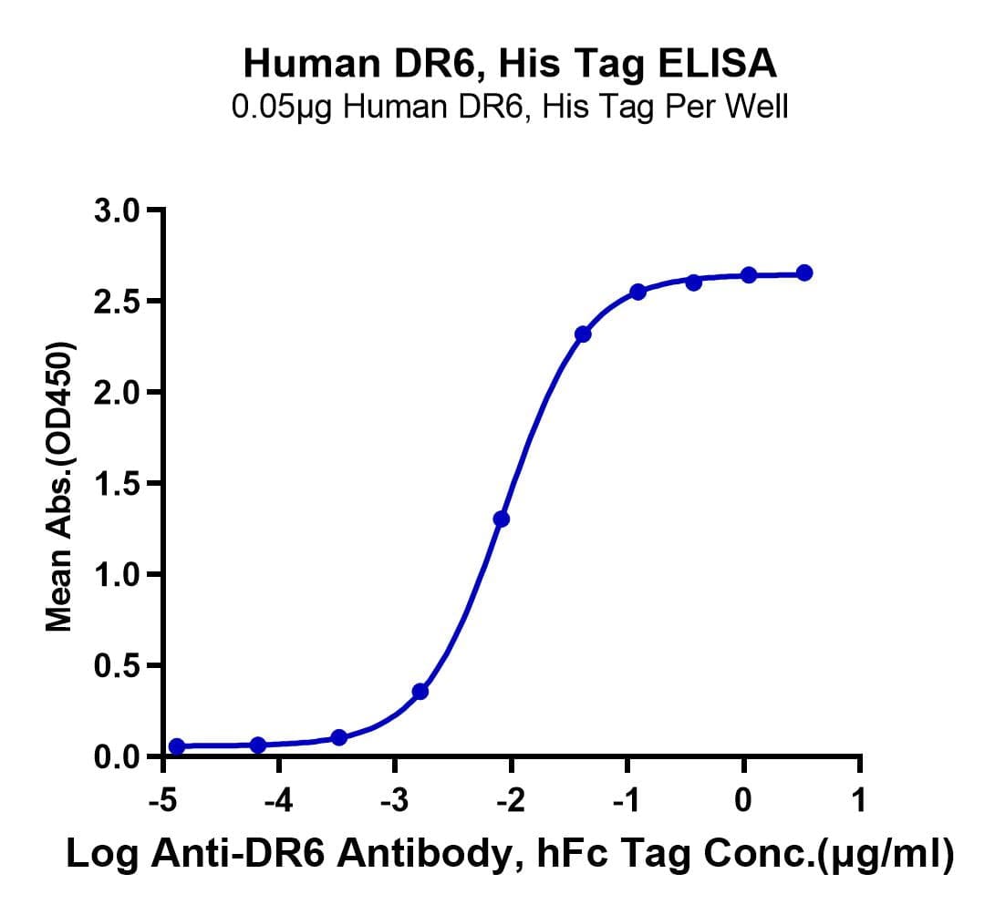
| Immobilized Human DR6, His Tag at 0.5µg/ml (100µl/Well) on the plate. Dose response curve for Anti-DR6 Antibody, hFc Tag with the EC50 of 8.7ng/ml determined by ELISA. |
|
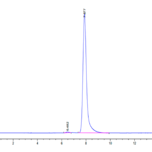
| Human DR6 on Tris-Bis PAGE under reduced condition. The purity is greater than 95%. |
Publications
Publications
| pmid | title | authors | citation |
|---|---|---|---|
| We haven't added any publications to our database yet. | |||
Protocols
| relevant to this product |
|---|
Documents
| # | ||
|---|---|---|
| Please enter your product and batch number here to retrieve product datasheet, SDS, and QC information. | ||