Notice: Function _load_textdomain_just_in_time was called incorrectly. Translation loading for the woocommerce-services domain was triggered too early. This is usually an indicator for some code in the plugin or theme running too early. Translations should be loaded at the init action or later. Please see Debugging in WordPress for more information. (This message was added in version 6.7.0.) in /www/benchmarkantibodiescom_769/public/wp-includes/functions.php on line 6121

Notice: Function _load_textdomain_just_in_time was called incorrectly. Translation loading for the woocommerce-payments domain was triggered too early. This is usually an indicator for some code in the plugin or theme running too early. Translations should be loaded at the init action or later. Please see Debugging in WordPress for more information. (This message was added in version 6.7.0.) in /www/benchmarkantibodiescom_769/public/wp-includes/functions.php on line 6121
Cytokeratin 7 – benchmark antibodies Cytokeratin 7
Skip to content

Showing all 2 results

Cytokeratin 7

Cytokeratin 7: Cytokeratin 7 is typically expressed in glandular epithelium and is used to identify adenocarcinomas of various origins, such as lung and ovarian cancers. Its detection aids in differentiating between primary and metastatic tumors, providing critical diagnostic information.


Human ovarian carcinoma stained with anti-Keratin 7 antibody using peroxidase-conjugate and DAB chromogen. Note the cytoplasmic staining of tumor cells.
IHC with antibody [6424] : Human ovarian carcinoma stained with anti-Keratin 7 antibody using peroxidase-conjugate and DAB chromogen. Note the cytoplasmic staining of tumor cells.
Human ovarian carcinoma stained with anti-Keratin 7 antibody using peroxidase-conjugate and DAB chromogen. Note the cytoplasmic staining of tumor cells.
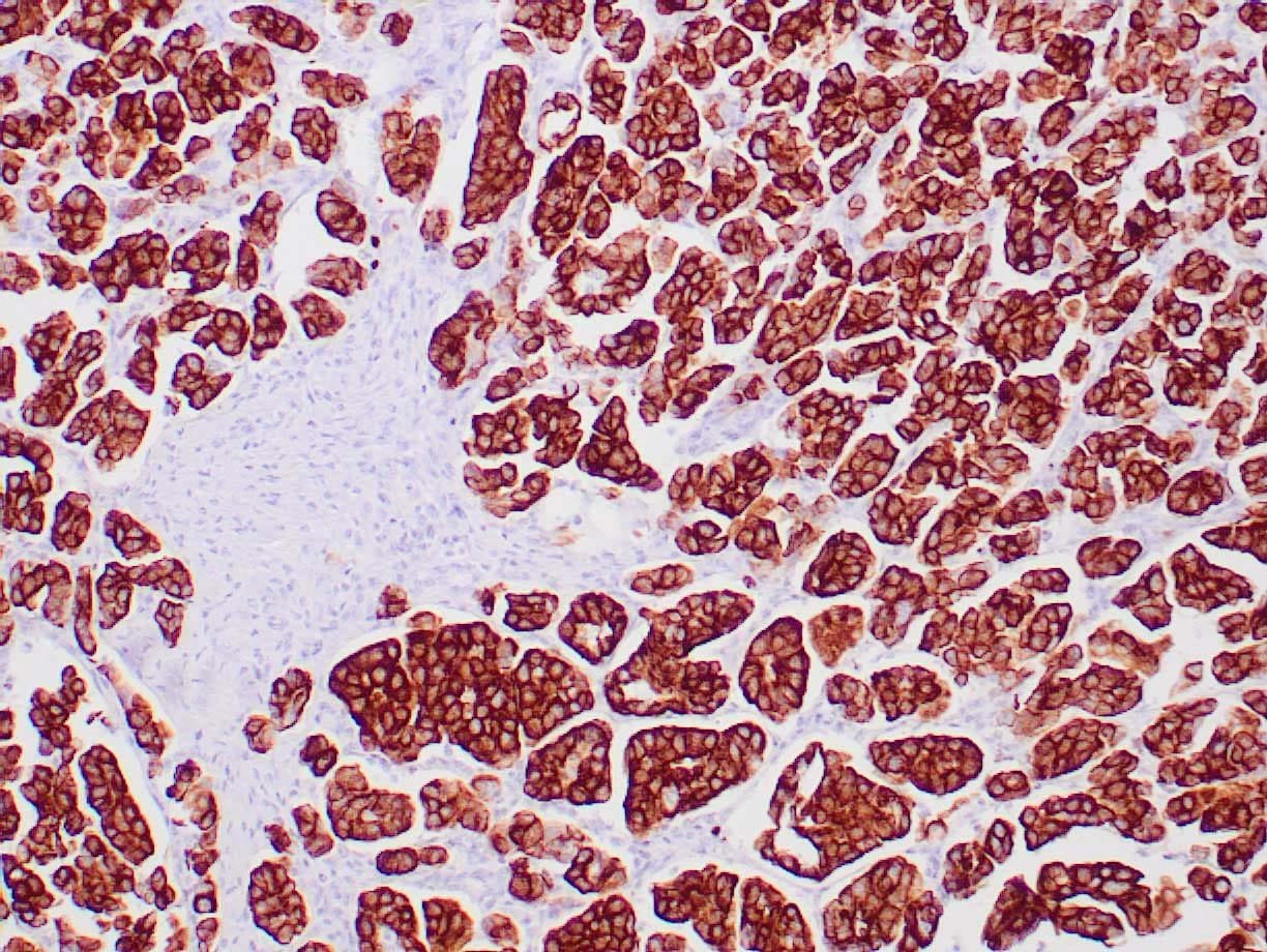
IHC with antibody [5423] : Human ovarian carcinoma stained with anti-Keratin 7 antibody using peroxidase-conjugate and DAB chromogen. Note the cytoplasmic staining of tumor cells.