Notice: Function _load_textdomain_just_in_time was called incorrectly. Translation loading for the woocommerce-services domain was triggered too early. This is usually an indicator for some code in the plugin or theme running too early. Translations should be loaded at the init action or later. Please see Debugging in WordPress for more information. (This message was added in version 6.7.0.) in /www/benchmarkantibodiescom_769/public/wp-includes/functions.php on line 6121

Notice: Function _load_textdomain_just_in_time was called incorrectly. Translation loading for the woocommerce-payments domain was triggered too early. This is usually an indicator for some code in the plugin or theme running too early. Translations should be loaded at the init action or later. Please see Debugging in WordPress for more information. (This message was added in version 6.7.0.) in /www/benchmarkantibodiescom_769/public/wp-includes/functions.php on line 6121
Cytokeratin 5/6 – benchmark antibodies Cytokeratin 5/6
Skip to content

Showing all 2 results

Cytokeratin 5/6

Cytokeratin 5/6: Cytokeratin 5/6 is often used to identify squamous cell carcinoma and is helpful in diagnosing mesotheliomas. Its expression is relevant in differentiating these tumors from other malignancies, aiding in accurate diagnosis and treatment planning.


Human prostate tissue stained with anti-Keratin 5/6 antibody using peroxidase-conjugate and DAB chromogen. Note the cytoplasmic staining of basal cells.
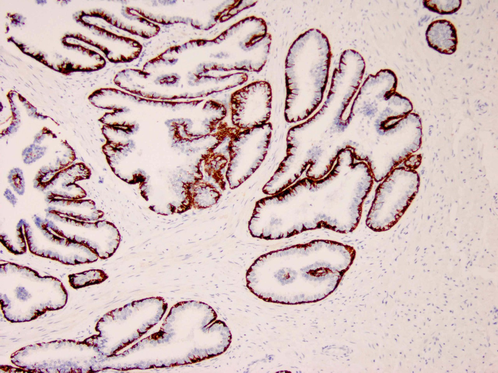
IHC with antibody [6147] : Human prostate tissue stained with anti-Keratin 5/6 antibody using peroxidase-conjugate and DAB chromogen. Note the cytoplasmic staining of basal cells.
Formalin-fixed, paraffin-embedded human prostate carcinoma stained with anti-cytokeratin 5/6 antibody using peroxidase-conjugate and DAB chromogen. Note the cytoplasmic staining of basal cells in the normal prostate (left), whereas carcinoma glands (right) are negative.
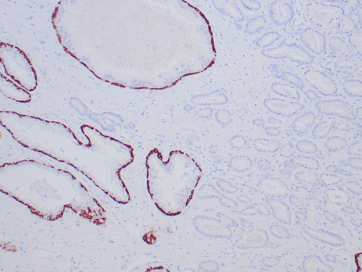
IHC with antibody [6148] : Formalin-fixed, paraffin-embedded human prostate carcinoma stained with anti-cytokeratin 5/6 antibody using peroxidase-conjugate and DAB chromogen. Note the cytoplasmic staining of basal cells in the normal prostate (left), whereas carcinoma glands (right) are negative.