Showing all 2 results
Cytokeratin 5
Cytokeratin 5: Cytokeratin 5 is primarily expressed in basal epithelial cells and is commonly used in diagnosing squamous cell carcinoma and mesothelioma. Its detection helps confirm the diagnosis of these tumors, providing valuable insights into tumor origin and differentiation.
| target | type | reactivity | applications | ||||
|---|---|---|---|---|---|---|---|
| 6145 | Cytokeratin 5 | rabbit monoclonal (ZR280) | human | IHC | |||
| 6146 | Cytokeratin 5 | mouse monoclonal (ZM186) | human | IHC | |||
| 6147 | Cytokeratin 5/6 | mouse monoclonal (D5&16B4) | human | IHC | |||
| 6148 | Cytokeratin 5/6 | rabbit monoclonal (ZR412) | human | IHC | |||
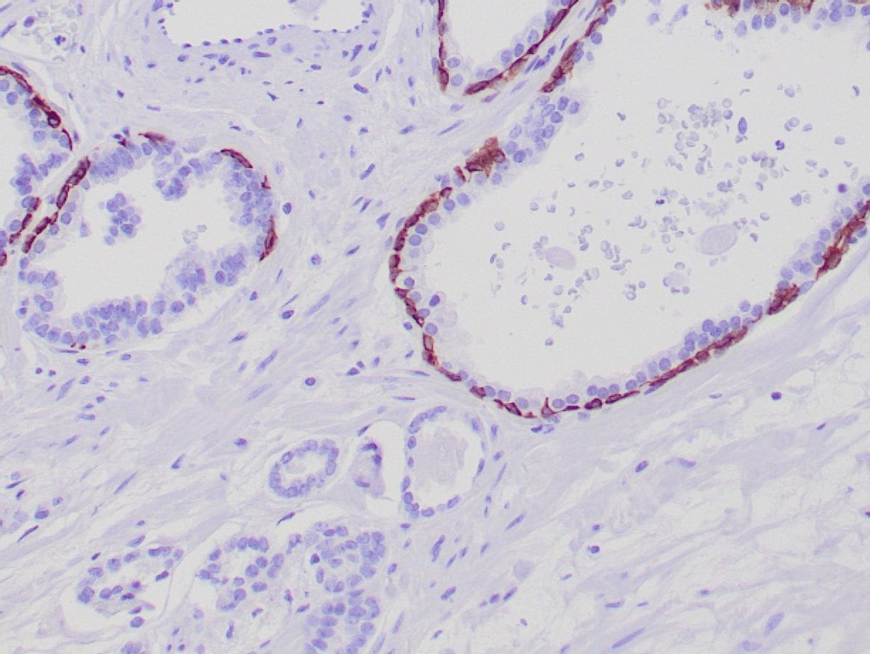
IHC with antibody [6145] : Formalin-fixed, paraffin-embedded human prostate stained with anti-keratin 5 antibody using peroxidase-conjugate and DAB chromogen. Note the cytoplasmic staining of basal cells in normal glands and negative stain in prostate carcinoma
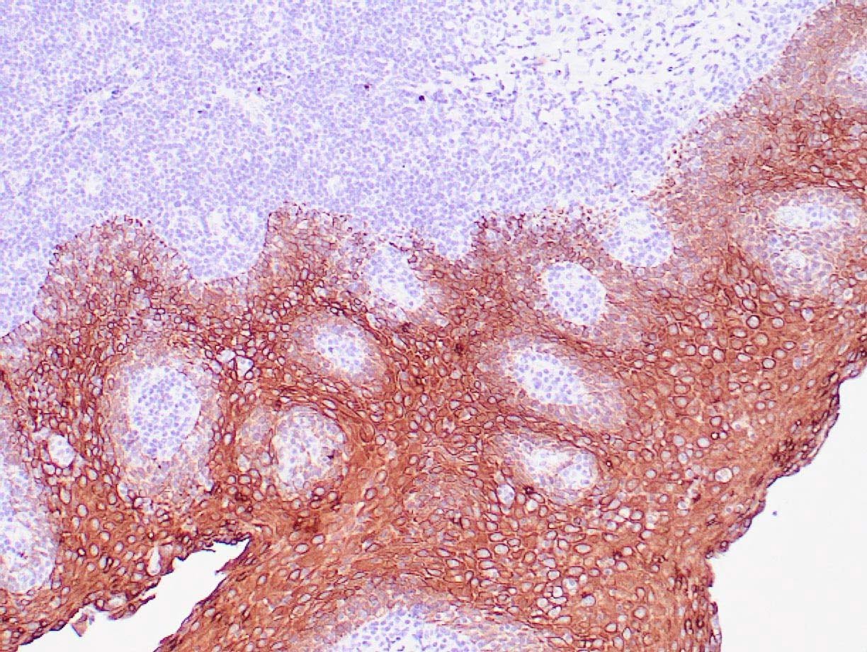
IHC with antibody [6146] : Human tonsil with anti-Keratin 5 antibody using peroxidase-conjugate and DAB chromogen. Note the cytoplasmic staining of squamous epithelium.

