t Human Helicobacter IgA Lateral flow dipstick kit 4775 – benchmark antibodies
no metadatahttps://benchmarkantibodies.com/product/human-helicobacter-iga-lateral-flow-dipstick-kit-4775/
Skip to content

Human Helicobacter IgA Lateral flow dipstick kit 4775

$487.00

Summary

  • Mikrogen diagnostik lateral flow device (dipstick) for research use (RUO)
  • Human Helicobacter IgA Lateral flow dipstick kit 4775
  • Suitable for IgA detection
  • Ready-to-use
  • 20 tests
SKU: 4775 Category: Tags: , ,
Weight1 lbs
Dimensions9 × 5 × 2 in
target

Human Helicobacter IgA

species reactivity

Human Helicobacter

applications

Lateral flow (dipstick)

assay type

Indirect & qualitative

available sizes

20 test kits

Human Helicobacter IgA Lateral flow dipstick kit 4775

kit
Assay type
Sandwich assay, lateral flow (dipstick)
Research area
Infectious Disease
Sample type
Serum, plasma, whole blood
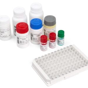
Components
10X Wash Buffer100 mL
TMB Substrate40 mL
Milk Powder5 g
Instructions for Use1 Each
Evaluation Form1 Each
Test Strips2 kits of [2 Vials x 10 Each]
Anti-Human IgA Conjugate500 μL
Positive Control140 μL
Negative Control140 μL
Storage
Store at 2-8°C.
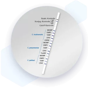
Additional information

Mikrogen recomLine Helicobacter 2.0 tests are serological, qualitative in vitro line immunoassays for the detection of antibodies against ten selected Helicobacter pylori antigens. The detection of antibodies against the two most important virulence factors CagA and VacA are of high diagnostic importance. These two virulence factors significantly increase the risk for premalignant changes, gastric carcinoma, MALT lymphoma, and ulcers. Humoral immune responses against the GroEL, HtrA, NapA, HP231, and CtkA biomarkers are associated with more severe disease progression.

Advantages
  • Sensitivity: > 99% IgG positive results of gold standard positive patients.
  • Specificity: 100% IgG and IgA negative results of gold standard negative patients.
  • Excellent correlation with the gold standard of Helicobacter pylori diagnostics - culture and histology.
  • Reliable results at all times, even with low colonization density.
  • Identification of highly virulent Helicobacter pylori type I infections using CagA.
  • Detection of antibodies against ten relevant biomarkers and virulence factors associated with more severe disease progression.
  • Simple and clear interpretation through easy-to-read banding.
  • Partial and full automation, software-based evaluation (recomScan), and integration with laboratory information system possible.
  • Highest sensitivity and specificity through the use of recombinant antigens:
Bands
AntigenDescriptionVirulence FactorSpecific for Type I / Type II
CagACytotoxin-associated gene AYesType I
VacAVaculating cytotoxin AYesType I / Type II
GroELChaperoneNoType I / Type II
FliDFlagella hook-associated proteinNoType I / Type II
HpaANeuraminyllactose-binding hemagglutinin, AdhesinYesType I / Type II
gGTGamma-Glutamyl TranspeptidaseYes<
target relevance
Organism
Helicobacter pylori
Structure and strains
Helicobacter pylori, previously known as Campylobacter pylori, is a gram-negative, flagellated, helical bacterium. Mutants can have a rod or curved rod shape, and these are less effective. Its helical body (from which the genus name, Helicobacter, derives) is thought to have evolved in order to penetrate the mucous lining of the stomach, helped by its flagella, and thereby establish infection. The bacterium was first identified as the causal agent of gastric ulcers in 1983 by the Australian doctors Barry Marshall and Robin Warren.
Disease
More than 50% of the world's population harbor Helicobacter pylori in their upper gastrointestinal tract. As a consequence, infections with this spirally formed, gram-negative bacterium belong to the most frequently occuring chronic bacterial diseases.

80 to 90% of all gastritis cases are traceable to an Helicobacter pylori infection. The person-to-person transmission is still not fully elucidated, but oral-oral and faecal-oral route mechanisms are discussed. Diseases associated with H. pylori infections include Ulcus duodeni, Ulcus ventriculi, stomach cancer and the seldom occuring MALT (Mucosa Associated Lymphatic Tissue) lymphoma. Phenotypic differences between different Helicobacter pylori isolates are limited to their ability to express the vacuolating cytotoxin (VacA) and its associated gene products (cytotoxin-associated genes; CagA). Due to phenotypic differences Helicobacter pylori isolates can be divided into virulent (type I) and non-virulent (type II) strains. Patients suffering from peptic or duodenal ulcers are more frequently infected with VacA and CagA producing Helicobacter pylori type I strains. Diagnose I
Detection and diagnosis
In the diagnosis of H. pylori infections, a distinction is made between non-invasive and invasive methods. Invasive procedures contain histology, urease rapid test and microbiological techniques such as cultivation and PCR. The C13-breath test, the Helicobacter pylori antigen detection in stool samples and serological antibody detection based on ELISA or immunoblot belong to the group of non-invasive methods. The detection of serum antibodies can be used for therapy control after eradication therapy

Publications

pmidtitleauthorscitation
We haven't added any publications to our database yet.
Published literature highly relevant to the biological target of this product and referencing this antibody or clone are retrieved from PubMed database provided by The United States National Library of Medicine at the National Institutes of Health.

relevant to this product
4775 protocol
#
Please enter your product and batch number here to retrieve product datasheet, SDS, and QC information.

Reviews

There are no reviews yet.

Only logged in customers who have purchased this product may leave a review.