| Weight | 1 lbs |
|---|---|
| Dimensions | 9 × 5 × 2 in |
| target | Echinococcus granulosus |
| species reactivity | Echinococcus (hydatid disease) |
| applications | ELISA |
| assay type | Indirect & quantitative |
| available size | 1 mg |
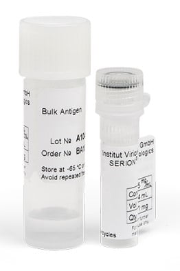
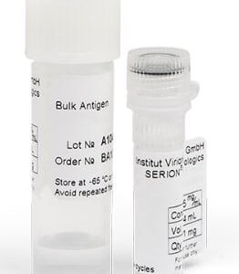
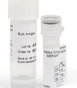
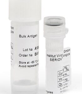
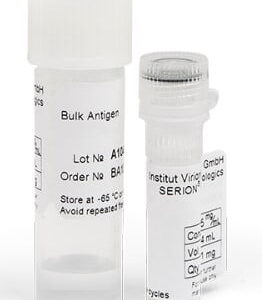
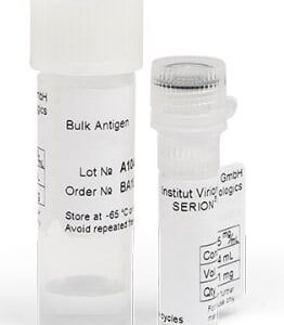
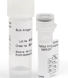
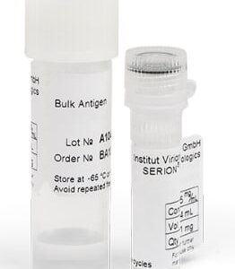
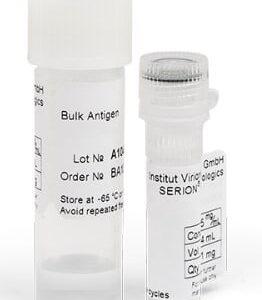
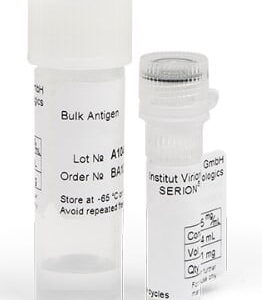
Echinococcus granulosus Antigen BA107VS
$741.00
Summary
- Virion/Serion Immunologics Antigen for research use (RUO)
- Echinococcus granulosus Antigen, recombinant
- Suitable for detection of IgA, IgG & IgM antibodies in ELISA
- Lot specific concentration, specified in mg/mL
- 1 mg
Echinococcus granulosus Antigen BA107VS
| kit |
|---|
| Research area Infectious Disease |
| Storage Store at -65°C or lower. Avoid repeated freeze-thaw cycles. 10 years from date of manufacture (under recommended storage conditions). |
| Form liquid |
| Associated products Echinococcus granulosus Antigen (BA107VS) Echinococcus IgG Control Serum (BC107G) Echinococcus IgG ELISA Kit (ESR107G) |
| target relevance |
|---|
| Organism Echinococcus granulosus |
| Protein names Echinococcus |
| Structure and strains Echinococcus granulosus, also called the hydatid worm, hyper tape-worm or dog tapeworm, is a cyclophyllid cestode that dwells in the small intestine of canids as an adult, but which has important intermediate hosts such as livestock and humans, where it causes cystic echinococcosis, also known as hydatid disease. The adult tapeworm ranges in length from 3 mm to 6 mm and has three proglottids ("segments") when intact an immature proglottid, mature proglottid and a gravid proglottid. The average number of eggs per gravid proglottid is 823. Like all cyclophyllideans, E. granulosus has four suckers on its scolex ("head"), and E. granulosus also has a rostellum with hooks. Several strains of E. granulosus have been identified, and all but two are noted to be infective in humans. |
| Detection and diagnosis Diagnosis of echinococcosis relies on clinical symptoms, imaging techniques (radiology, computer tomography) as well as on serological investigations. |
Data
Publications
| pmid | title | authors | citation |
|---|---|---|---|
| We haven't added any publications to our database yet. | |||
Protocols
| relevant to this product |
|---|
| BA107VS protocol |
Documents
| Product data sheet |
|---|
| BA107VS |
Only logged in customers who have purchased this product may leave a review.
Reviews
There are no reviews yet.