Showing the single result
HSP60 antibodies
Heat shock protein 60 (HSP60) is a mitochondrial chaperone protein that plays a pivotal role in protein folding, assembly, and stabilization within cells. It is part of the cell's stress response system, ensuring proper protein conformation and preventing misfolding, particularly under stressful conditions such as heat shock, oxidative stress, or infection. HSP60 is essential for maintaining mitochondrial function and overall cellular homeostasis, and it is involved in various processes, including apoptosis and immune response.
In Western blotting (WB), HSP60 is widely used to monitor mitochondrial integrity and the cellular response to stress. The HSP60 antibodies offered on your website can also be applied in immunofluorescence (IF) and immunohistochemistry (IHC) to visualize its localization in mitochondria and other compartments. This is particularly useful in research focused on mitochondrial dysfunction, neurodegenerative diseases, cancer, and metabolic disorders, where HSP60 levels often fluctuate in response to cellular damage or stress.
Given its critical role in protein maintenance and stress response, HSP60 is an indispensable marker in studies investigating mitochondrial health, cellular stress mechanisms, and protein homeostasis, providing insights into various physiological and pathological conditions.
| target | type | reactivity | applications | ||||
|---|---|---|---|---|---|---|---|
| 9023 | Hsp60 | rabbit polyclonal | human mouse rat | WB ICC IHC | |||
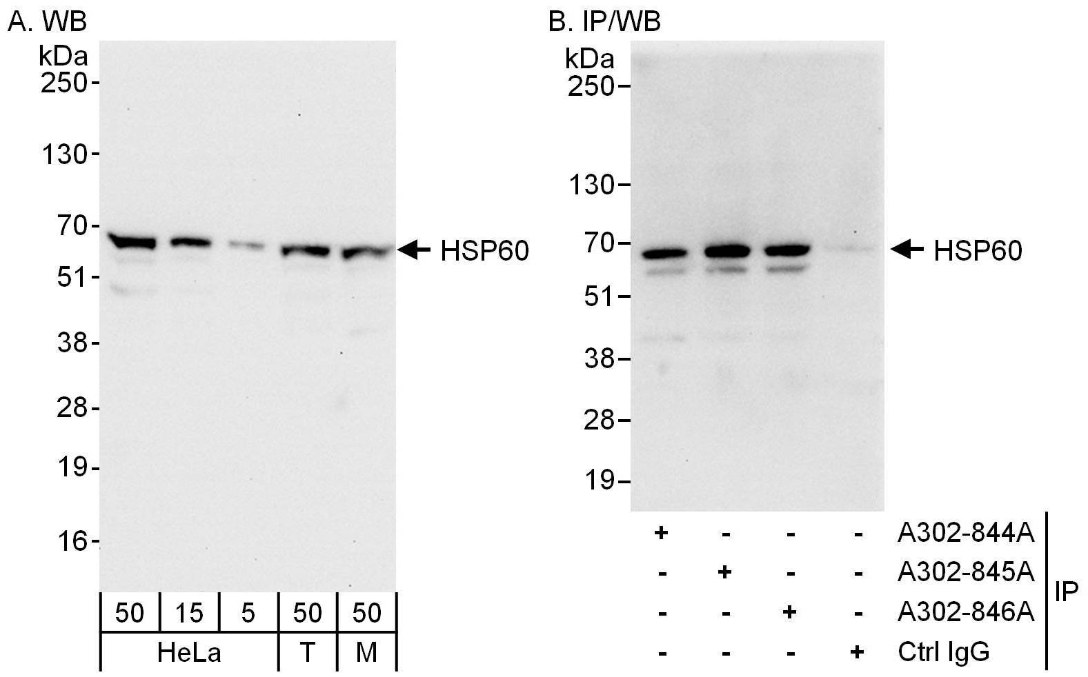
WB with antibody [9023] : Detection of human and mouse HSP60 by western blot (h&m) and immunoprecipitation (h).
Samples: Whole cell lysate from HeLa (5, 15 and 50 �g for WB; 1 mg for IP, 20% of IP loaded), HEK293T (T; 50 �g), and mouse NIH 3T3 (M; 50 �g) cells. Antibodies: Affinity purified rabbit anti-HSP60 antibody used for WB at 0.04 �g/ml (A) and 0.4 �g/ml (B) and used for IP at 3 �g/mg lysate. HSP60 was also immunoprecipitated by rabbit anti-HSP60 antibodies, which recognize other epitopes. Detection: Chemiluminescence with exposure times of 10 seconds (A and B).

IHC with antibody [9023] : Detection of human HSP60 by immunohistochemistry.
Sample: FFPE section of human ovarian carcinoma. Antibody: Affinity purified rabbit anti-HSP60 used at a dilution of 1:200 (1�g/ml). Detection: Vector Laboratories ImmPACT NovaRED Peroxidase Substrate
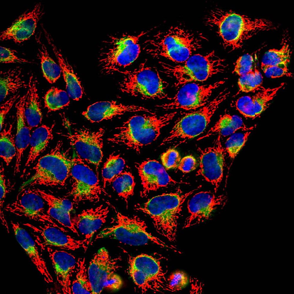
ICC/IF with antibody [2417] : Confocal immunofluorescent analysis of HeLa cells stained with rabbit pAb to HSP60, 2417, dilution 1:1,000, in red, and costained with chicken pAb to vimentindilution 1:1,000 in green. The blue is DAPI staining of nuclear DNA. The HSP60 antibody gives strong and specific staining of mitochondria while the vimentin antibody reveals cytoplasmic intermediate filaments.
