Showing all 2 results
GAPDH antibodies
Glyceraldehyde 3-phosphate dehydrogenase (GAPDH) is a key enzyme in glycolysis, responsible for catalyzing the conversion of glyceraldehyde 3-phosphate into 1,3-bisphosphoglycerate, an essential step in cellular energy production. Beyond its critical metabolic role, GAPDH has become a widely used reference protein in molecular biology and biochemistry research. Its stable expression across a range of cell types and conditions makes it an ideal loading control in Western blotting (WB) and gel electrophoresis.
As a loading control, GAPDH helps normalize protein levels in experimental samples, ensuring accurate quantification and comparison of target proteins by compensating for variations in protein loading and gel transfer efficiency. The antibodies for GAPDH offered on your website are also suitable for immunofluorescence (IF) and immunohistochemistry (IHC) applications, enabling visualization of its cellular localization alongside metabolic pathways. Its consistent expression, both under normal and experimental conditions, provides a reliable baseline for comparative analysis, making GAPDH an indispensable tool for researchers studying cellular processes and protein expression levels.
| target | type | reactivity | applications | ||||
|---|---|---|---|---|---|---|---|
| 1937 | GAPDH | mouse monoclonal (GA1R) | human mouse rat rabbit hamster | WB ICC ELISA | |||
| 9013 | GAPDH | rabbit polyclonal | human mouse rat | WB ICC IHC | |||

WB with antibody [1937] : Three loading control mAbs reacting against 10µg/lane of mouse brain tissue lysates. 50kDa band is Anti-β-Tubulin (#5007) at 1:2,000 dilution (0.5 µg/mL); 42kDa band is Anti-β-Actin (#5132) at 1:1,000 dilution (1 µg/mL); 37kDa band is Anti-GAPDH (#1937) at 1:5,000 dilution (0.2 µg/mL).

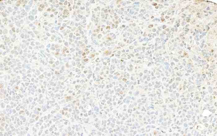
IHC with antibody [9013] : Detection of mouse GAPDH by immunohistochemistry. Sample: FFPE section of mouse plasmacytoma. Antibody: Affinity purified rabbit anti-GAPDH antibody used at a dilution of 1:1000 (0.2µg/ml). Detection: DAB

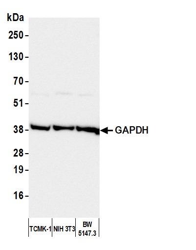
WB with antibody [9013] : Detection of mouse GAPDH by western blot.
Samples: Whole cell lysate (50 µg) from TCMK-1, NIH 3T3, and BW5147.3 cells prepared using NETN lysis buffer. Antibody: Affinity purified rabbit anti-GAPDH antibody used for WB at 0.04 µg/ml. Detection: Chemiluminescence with an exposure time of 3 seconds.


