Notice: Function _load_textdomain_just_in_time was called incorrectly. Translation loading for the woocommerce-services domain was triggered too early. This is usually an indicator for some code in the plugin or theme running too early. Translations should be loaded at the init action or later. Please see Debugging in WordPress for more information. (This message was added in version 6.7.0.) in /www/benchmarkantibodiescom_769/public/wp-includes/functions.php on line 6121

Notice: Function _load_textdomain_just_in_time was called incorrectly. Translation loading for the woocommerce-payments domain was triggered too early. This is usually an indicator for some code in the plugin or theme running too early. Translations should be loaded at the init action or later. Please see Debugging in WordPress for more information. (This message was added in version 6.7.0.) in /www/benchmarkantibodiescom_769/public/wp-includes/functions.php on line 6121
CD3 – benchmark antibodies CD3
Skip to content

Showing all 2 results

CD3

CD3: CD3 is a pan-T-cell marker and is critical for assessing T-cell populations in tumors. Its expression is relevant in diagnosing lymphomas and understanding the immune landscape in various cancers, aiding in treatment decisions.


target type reactivity applications
No products found

Formalin-fixed, paraffin-embedded human lymph node stained with anti-CD3 antibody using peroxidase-conjugate and DAB chromogen. Note the membranous staining of perifollicular T-cells and no stain in B-cells
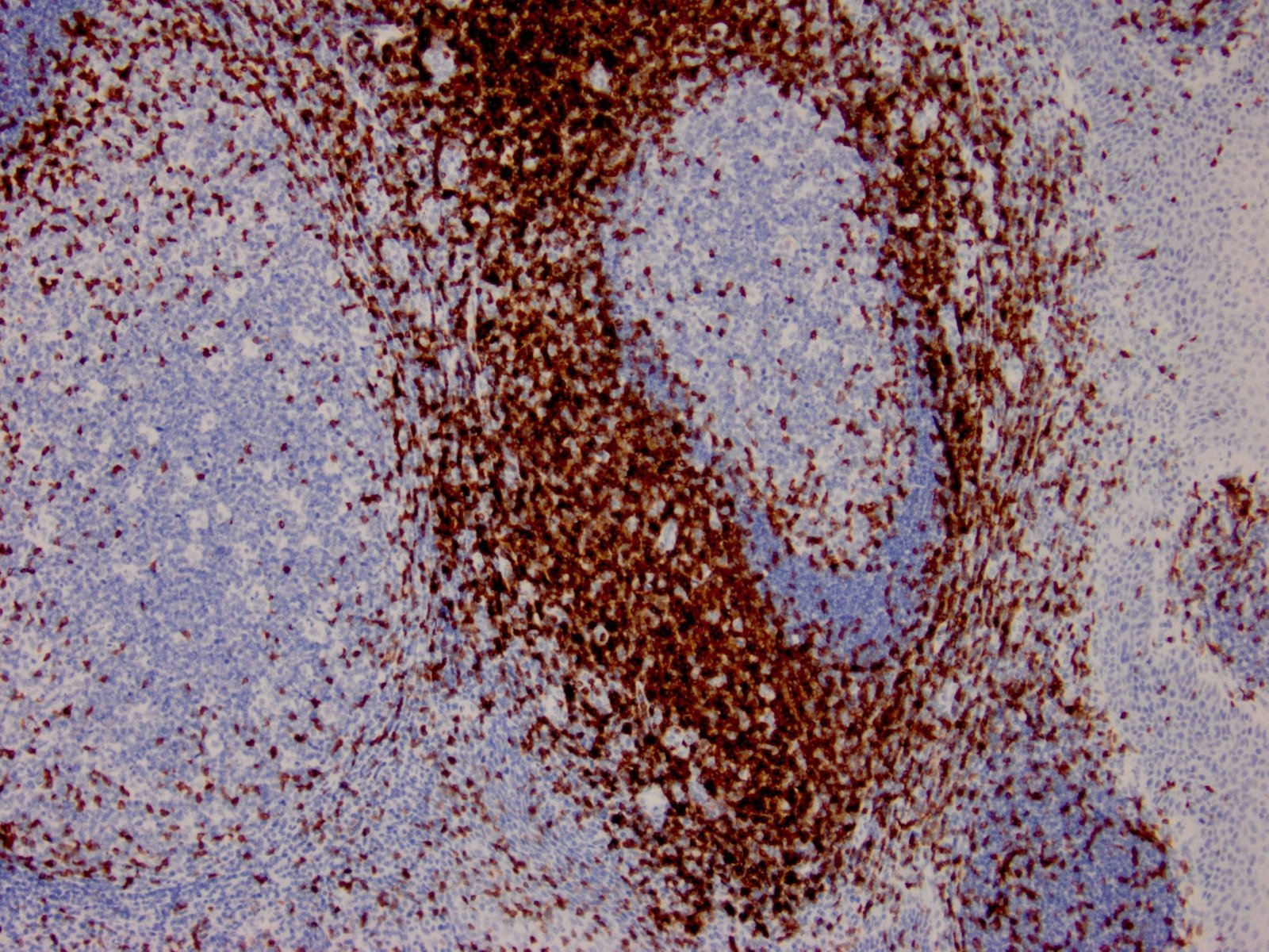
IHC with antibody [6081] : Formalin-fixed, paraffin-embedded human lymph node stained with anti-CD3 antibody using peroxidase-conjugate and DAB chromogen. Note the membranous staining of perifollicular T-cells and no stain in B-cells
Human lymph node stained with anti-CD3 antibody using peroxidase-conjugate and DAB chromogen. Note the membranous staining of perifollicular T-cells and no stain in B-cells.
IHC with antibody [6421] : Human lymph node stained with anti-CD3 antibody using peroxidase-conjugate and DAB chromogen. Note the membranous staining of perifollicular T-cells and no stain in B-cells.