Showing the single result
Calreticulin antibodies
Calreticulin is a multifunctional calcium-binding chaperone protein primarily located in the endoplasmic reticulum (ER), where it plays a critical role in protein folding, quality control, and calcium homeostasis. It assists in the proper folding of newly synthesized glycoproteins and ensures that only correctly folded proteins exit the ER. Calreticulin is also involved in immune response modulation and cell adhesion processes, making it relevant in various physiological and pathological contexts, including cancer, autoimmune diseases, and cardiac development.
In Western blotting (WB), calreticulin is commonly used as a marker for ER stress and protein-folding capacity. The calreticulin antibodies offered on your website are also suitable for immunofluorescence (IF) and immunohistochemistry (IHC), allowing researchers to visualize its expression and localization in the ER and other compartments within cells. This helps investigate ER stress, apoptosis, and immune-related pathways, particularly in cancer and inflammatory diseases, where calreticulin exposure on the cell surface can trigger immune recognition.
Given its essential role in ER function, protein folding, and calcium regulation, calreticulin is a valuable marker in studies focusing on cellular stress, immune modulation, and protein quality control. It is especially important in research on cancer, autoimmunity, and cardiovascular diseases, where its expression and function are often dysregulated.
| target | type | reactivity | applications | ||||
|---|---|---|---|---|---|---|---|
| 9015 | Calreticulin | rabbit polyclonal | human mouse rat | WB ICC IHC | |||
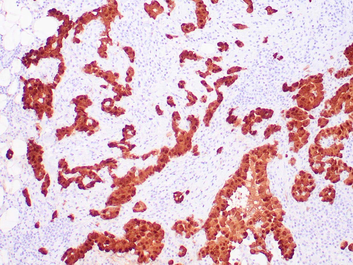
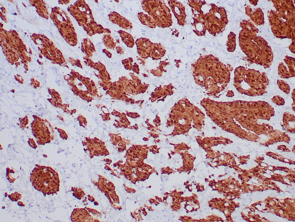
IHC with antibody [6052] : Human mesothelioma stained with anti-calretinin antibody using peroxidase-conjugate and DAB chromogen. Note nuclear/cytoplasmic staining of mesothelioma cells.
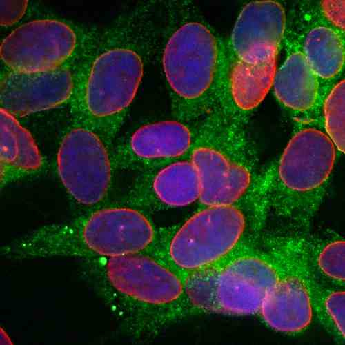
ICC/IF with [1726] : Immunofluorescent analysis of of SH-SY5Y cells stained with mouse mAb to calreticulin, MCA-6C6, dilution 1:500 in green and costained with chicken pAb to lamin A/C, CPCA-LaminAC, dilution 1:2,000 in red. The blue is DAPI staining of nuclear DNA. The MCA-6C6 antibody reveals granular staining of cytoplasm, while the lamin A/C antibody stains the nuclear lamina and membrane.
