Immunohistochemistry (IHC) is a powerful technique that allows scientists and researchers to visualize and analyze the distribution of specific proteins or antigens within tissue samples. One of the key steps in the IHC process is cell staining, where various staining methods are employed to enhance contrast and highlight specific cellular components. In this blog post, we will delve into the diverse world of cell staining techniques used in immunohistochemistry, shedding light on their principles and applications.
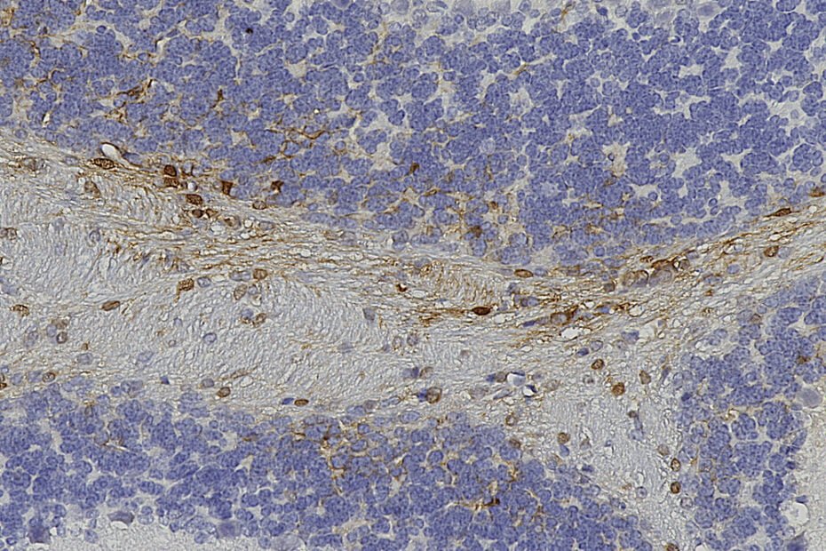
The most common type of cell staining in immunohistochemistry involves the use of chromogens, substances that produce a visible color change when they react with specific target molecules. The peroxidase, HRP, substrate diaminobenzidine (DAB) is a widely used chromogen that yields a brown color, making it easy to identify the presence and localization of target antigens under a microscope. This technique is especially valuable in identifying the spatial distribution of proteins within tissues and understanding cellular function.
Enzyme-based cell staining methods contribute significantly to the versatility of immunohistochemistry. Alkaline phosphatase (AP) and horseradish peroxidase (HRP) are commonly employed enzymes that catalyze color-producing reactions. These enzymes can be conjugated to antibodies, enabling the visualization of target antigens. The choice between AP and HRP depends on factors such as substrate availability, sensitivity, and desired color output.
Immunohistochemistry also embraces non-enzymatic special stains that offer unique insights into cellular structures and functions. Hematoxylin and eosin (H&E) staining, for instance, is a routine technique that imparts a purple-blue color to cell nuclei (hematoxylin) and a pink color to cytoplasmic structures (eosin). While not specific to immunohistochemistry, H&E staining remains a fundamental method for assessing tissue morphology and is often used in conjunction with IHC for comprehensive tissue analysis.
Fluorescent cell staining represents another emerging dimension of immunohistochemistry. Unlike chromogenic stains, fluorescent dyes emit light of various colors when exposed to specific wavelengths. This allows for the simultaneous visualization of multiple antigens within the same tissue section, enabling researchers to study co-localization and interactions between different proteins. Fluorescent staining is not only visually appealing but also provides quantitative data through image analysis, making it a valuable tool in modern biomedical research.
In recent years, advancements in technology have given rise to automated staining platforms, streamlining the immunohistochemistry workflow. These platforms not only enhance the reproducibility of results but also allow for high-throughput analysis, making them particularly beneficial in large-scale research studies and clinical settings. Automated systems ensure consistent and standardized staining procedures, reducing variability and improving the reliability of experimental outcomes.
In conclusion, the world of cell staining in immunohistochemistry is rich and diverse, offering a spectrum of techniques to cater to the specific needs of researchers. Whether using chromogens, fluorescent dyes, special stains, enzyme-based methods, or automated platforms, each staining technique brings its own advantages to the table. By harnessing the power of these techniques, scientists can unravel the intricate details of cellular structures and functions, advancing our understanding of disease mechanisms and contributing to the progress of biomedical research.
