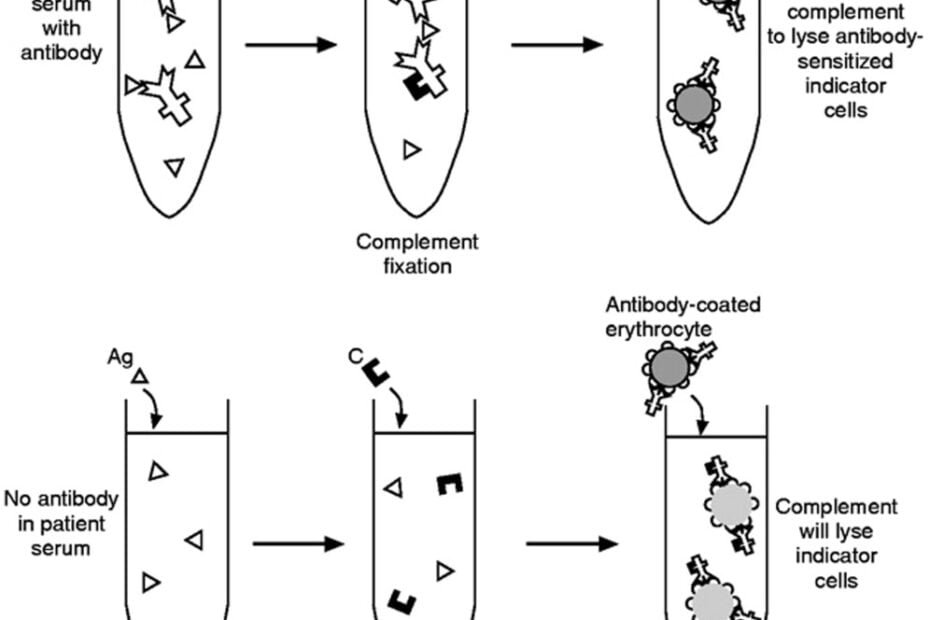
Introduction:
In the realm of immunological testing, the Complement Fixation Test (CFT) stands as a stalwart method, offering insights into various infectious and autoimmune diseases. Understanding how this assay is performed and the materials needed is crucial for appreciating its significance in medical diagnostics. In this article, we delve into the procedural intricacies of the CFT and the essential materials required for its execution.
Procedure Overview:
The Complement Fixation Test operates on the principle of antigen-antibody interaction and the subsequent activation of the complement system. Here’s a simplified overview of the procedure:
- Serum Collection: The first step involves obtaining a serum sample from the patient suspected of having the target disease.
- Preparation of Antigen: Known antigens specific to the suspected pathogen or antigen of interest are prepared. These antigens can be obtained from cultured microorganisms or synthesized in the laboratory.
- Serum Dilution: The patient’s serum is diluted to various concentrations to ensure that the test is sensitive enough to detect the presence of antibodies.
- Mixing Serum and Antigen: Diluted serum samples are mixed with the prepared antigen. If antibodies against the antigen are present in the serum, they will bind to the antigen molecules, forming antigen-antibody complexes.
- Addition of Complement Proteins: Complement proteins are added to the mixture. If the complement proteins become fixed (bound) to the antigen-antibody complexes, it indicates a positive reaction.
- Indicator System: An indicator system is employed to visualize the results of complement fixation. This system can involve various methods, such as the use of indicator cells or detecting a change in turbidity or color.
Materials Needed:
Executing the Complement Fixation Test requires a set of specific materials to ensure accuracy and reliability. Here’s a list of essential components:
- Serum Samples: Serum samples obtained from patients suspected of having the target disease are necessary for testing.
- Antigens: Specific antigens relevant to the disease being investigated are required. These antigens can be obtained commercially or prepared in the laboratory.
- Complement Proteins: Fresh or preserved complement proteins are essential for the assay. Complement proteins can be sourced from animal sera or obtained commercially.
- Indicator System: Depending on the method chosen for visualizing complement fixation, various indicator systems may be employed. This can include indicator cells (sheep or guinea pig red blood cells), hemolytic systems, or indicators for detecting changes in turbidity or color.
- Laboratory Equipment: Standard laboratory equipment such as microscopes, centrifuges, pipettes, and incubators are necessary for conducting the assay.
- Reagents and Buffers: Reagents for diluting serum samples, preparing antigens, and maintaining the stability of complement proteins are essential. Additionally, buffers to maintain pH levels and optimize reaction conditions are required.
Conclusion:
The Complement Fixation Test remains a valuable tool in immunological testing, providing insights into a wide range of infectious and autoimmune diseases. Understanding the procedural steps and materials required for its execution is essential for accurate and reliable results. By appreciating the intricacies of the CFT, healthcare professionals can leverage this assay effectively in diagnosing and monitoring various medical conditions, ultimately improving patient care and outcomes.
