|
Protocol - FLAG-Tag (DYKDDDDK-Tag Proteins) ELISA
Catalog #: 60811
For the quantitative measurement of FLAG-Tag (DYKDDDDK-Tag Proteins) in supernatants.
|
|
FLAG-Tag (DYKDDDDK-Tag Protein) ELISA Kit
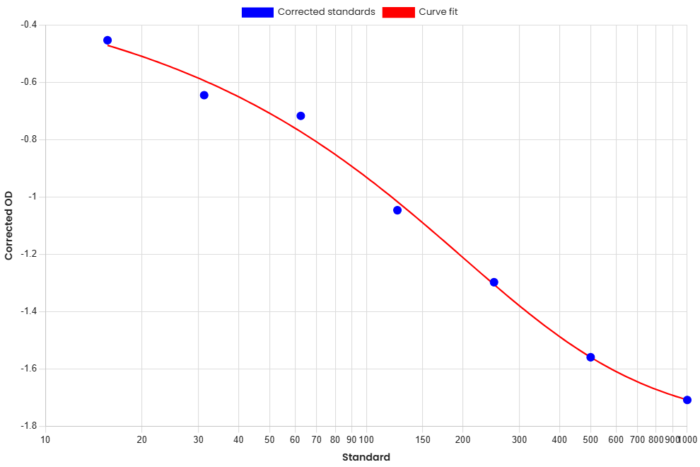
This ELISA is a solid-phase competiton enzyme-linked immunosorbent assay (cELISA) for the quantitative measurement of FLAG-Tag (DYKDDDDK-Tag Proteins) in biological samples. This assay is highly specific and sensitive, providing a reliable method for quantitative analysis.
This assay employs a competitive ELISA for the quantitative measurement of anti-FLAG-Tag (DYKDDDDK-Tag Proteins) antibodies. Samples and Standards are mixed with 1X Biotinylated Detector Reagent and added to antigen-coated microplate wells. Anti-FLAG-Tag (DYKDDDDK-Tag Proteins) antibodies present in the sample compete with the biotinylated detector reagent for binding to the immobilized antigen. Following incubation, unbound material is removed by washing. 1X HRP-Streptavidin Reagent is then added to bind the biotinylated detector reagent remaining on the plate. After a further wash, TMB Substrate Solution is added, producing a colorimetric signal that is inversely proportional to the concentration of anti-FLAG-Tag (DYKDDDDK-Tag Proteins) antibodies in the sample. The reaction is terminated by the addition of Stop Solution, and absorbance is measured. Anti-FLAG-Tag (DYKDDDDK-Tag Proteins) antibody concentrations are determined by comparison with the Standard Curve.
| Component | Description |
|---|---|
| ELISA Microplate | 96-well (12 strips × 8) coated microplate |
| Lyophilized Standard | 2 vials |
| Biotinylated Detector Reagent (100X) | 60 µL |
| HRP-Streptavidin Reagent (100X) | 120 µL |
| Sample Dilution Buffer | 20 mL |
| Detector Dilution Buffer | 10 mL |
| HRP-Streptavidin Dilution Buffer | 10 mL |
| Wash Buffer Concentrate (25X) | 30 mL |
| Substrate Solution (TMB) | 10 mL |
| Stop Solution | 10 mL |
| Plate Sealers | 5 adhesive sealing films |
* Store the unopened kit at 2–8°C. Do not use past the expiration date. Do not combine materials from different lots.
The following are general guidelines. Sample stability has not been fully characterized.
Bring all reagents to room temperature before use. 1X Wash Buffer may be prepared in full at any time. Prepare only the required volume of 1X Biotinylated Detector Reagent and 1X HRP-Streptavidin Reagent, no more than 30 minutes before use.
Prepare diluted samples and the Standard dilution series. Use all diluted Standards and samples within 2 hours. A second vial of Lyophilized Standard is provided for later experiments.
| 1. | Bring all reagents and samples to room temperature. Prepare Standards, 1X Wash Buffer, 1X Biotinylated Detector Reagent, 1X HRP-Streptavidin Reagent, and appropriately diluted samples. Place the required number of microplate strips into the plate frame. Duplicate Standard and sample measurements are recommended. Return unused strips to the foil pouch containing the desiccant pack, and reseal. |
| 2. | Wash the plate wells twice before adding standards and samples. Add 50 µL of each Standard to the designated wells. Add 50 µL of each sample to the designated wells. Immediately add Add 50 µL of 1X Biotinylated Detector Reagent to all wells. Tap plate gently to mix. Apply a plate sealer and incubate at 37°C for 45 minutes. Record sample positions. |
| 3. | Aspirate the contents of each well and wash three times with 350 µL of 1X Wash Buffer per wash. Ensure complete removal of liquid at each step. After the last wash, remove any remaining Wash Buffer by inverting the plate and blotting it against clean paper towels. |
| 4. | Add 100 µL of 1X HRP-Streptavidin Reagent to all wells. Apply a plate sealer and incubate at 37°C for 30 minutes. |
| 5. | Aspirate the contents of each well and wash five times with 350 µL of 1X Wash Buffer per wash. Wash and blot the plate as described above. |
| 6. | Add 90 µL of Substrate Solution (TMB) to all wells. Incubate at 37°C, protected from direct light, for 10–20 minutes. Monitor color development. The reaction may be stopped when an appreciable color gradient is observed across the Standard dilution series. |
| 7. | Add 50 µL of Stop Solution to each well. The color in the wells should change from blue to yellow. Gently tap the plate to mix the contents of each well. |
| 8. | Measure the Optical Density (OD) of each well within 30 minutes using a microplate reader set to 450 nm. If wavelength correction is available, set correction to 540 nm or 570 nm. |
| Standard | Amount [ng/mL] | OD1 | OD2 | Average | Corrected |
|---|---|---|---|---|---|
| Blank #8 | 0 | 2.28 | 2.39 | 2.32 | 2.32 |
| Standard #7 | 15.63 | 1.84 | 1.93 | 1.87 | 1.87 |
| Standard #6 | 31.25 | 1.65 | 1.73 | 1.68 | 1.68 |
| Standard #5 | 62.5 | 1.58 | 1.66 | 1.61 | 1.61 |
| Standard #4 | 125 | 1.26 | 1.32 | 1.28 | 1.28 |
| Standard #3 | 250 | 1.01 | 1.07 | 1.03 | 1.03 |
| Standard #2 | 500 | 0.76 | 0.8 | 0.77 | 0.77 |
| Standard #1 | 1000 | 0.61 | 0.64 | 0.63 | 0.63 |
| Sample | n | Mean | Standard Deviation | CV (%) |
|---|---|---|---|---|
| Intra Sample 1 | 20 | 30.63 | 1.38 | 4.51 |
| Inter Sample 1 | 20 | 31.16 | 1.4 | 4.48 |
| Intra Sample 2 | 20 | 121.9 | 6.02 | 4.94 |
| Inter Sample 2 | 20 | 125.3 | 6.01 | 4.8 |
| Intra Sample 3 | 20 | 518 | 25.23 | 4.87 |
| Inter Sample 3 | 20 | 523 | 23.22 | 4.44 |
| Matrix | Recovery Range (%) | Average (%) |
|---|---|---|
| Cell Culture Lysate(n=5) | 85-105 | 95 |
| Sample | 1:2 | 1:4 | 1:8 |
|---|---|---|---|
| Cell Culture Lysate(n=5) | 82-98% | 84-101% | 90-98% |
| 37°C / 1 Month | 2-8°C / 6 Months | 2-8°C / 12 Months |
|---|---|---|
| 80 | 95-100 | 85-98 |