VERO host cell proteins (HCP) ELISA Kit Protocol - 60807
Protocol - VERO host cell proteins (HCP) sandwich ELISA
Catalog #: 60807
For the quantitative measurement of VERO host cell proteins (HCP) in supernatants.
Benchmark Antibodies Logo
For research use only. Not for use in diagnostic procedures.
Introduction


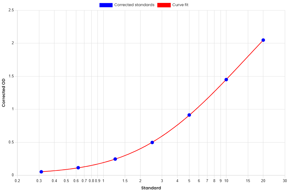
This ELISA is a solid-phase sandwich enzyme-linked immunosorbent assay (sELISA) for the quantitative measurement of VERO host cell proteins (HCP) in biological samples. This assay is highly specific and sensitive, providing a reliable method for quantitative analysis.

Principle of the Assay

This assay employs an antibody sandwich ELISA for the measurement of VERO host cell proteins (HCP). Samples and Standards are incubated in anti-host cell proteins (HCP) antibody coated wells, followed by 1X Biotinylated Detector Reagent Antibody and 1X HRP-Streptavidin Reagent. After washing, Substrate Solution (TMB) produces a colorimetric reaction proportional to the amount of bound host cell proteins (HCP). Following addition of Stop Solution, absorbance is measured and host cell proteins (HCP) concentrations are calculated from the Standard Curve.

Materials Provided & Storage Conditions
Component Description
ELISA Microplate 96-well (12 strips × 8) coated microplate
Lyophilized Standard 2 vials
Biotinylated Detector Reagent (100X) 120 µL
HRP-Streptavidin Reagent (100X) 120 µL
Sample Dilution Buffer 20 mL
Detector Dilution Buffer 20 mL
HRP-Streptavidin Dilution Buffer 10 mL
Wash Buffer Concentrate (25X) 30 mL
Substrate Solution (TMB) 10 mL
Stop Solution 10 mL
Plate Sealers 5 adhesive sealing films

* Store the unopened kit at 2–8°C. Do not use past the expiration date. Do not combine materials from different lots.

Other Supplies Required
Precautions & Technical Hints
Sample Collection & Storage

The following are general guidelines. Sample stability has not been fully characterized.

Reagent Preparation

Bring all reagents to room temperature before use. 1X Wash Buffer may be prepared in full at any time. Prepare only the required volume of 1X Biotinylated Detector Reagent and 1X HRP-Streptavidin Reagent, no more than 30 minutes before use.

Sample and Standards Preparation

Prepare diluted samples and the Standard dilution series. Use all diluted Standards and samples within 2 hours. A second vial of Lyophilized Standard is provided for later experiments.

Assay Procedure
1.Bring all reagents and samples to room temperature. Prepare Standards, 1X Wash Buffer, 1X Biotinylated Detector Reagent, 1X HRP-Streptavidin Reagent, and appropriately diluted samples. Place the required number of microplate strips into the plate frame. Duplicate Standard and sample measurements are recommended. Return unused strips to the foil pouch containing the desiccant pack, and reseal.
2.Add 100 µL of each Standard to the designated wells. Add 100 µL of each sample to the designated wells. Record sample positions. Apply a plate sealer and incubate at 37°C for 90 minutes.
3.Aspirate the contents of each well and wash twice with 350 µL of 1X Wash Buffer per wash. Ensure complete removal of liquid at each step. After the last wash, remove any remaining Wash Buffer by inverting the plate and blotting it against clean paper towels.
4.Add 100 µL of 1X Biotinylated Detector Reagent to all wells. Apply a plate sealer and incubate at 37°C for 60 minutes.
5.Aspirate the contents of each well and wash three times with 350 µL of 1X Wash Buffer per wash. Wash and blot the plate as described above.
6.Add 100 µL of 1X HRP-Streptavidin Reagent to all wells. Apply a plate sealer and incubate at 37°C for 30 minutes.
7.Aspirate the contents of each well and wash five times with 350 µL of 1X Wash Buffer per wash. Wash and blot the plate as described above.
8.Add 90 µL of Substrate Solution (TMB) to all wells. Incubate at 37°C, protected from direct light, for 10–20 minutes. Monitor color development. The reaction may be stopped when an appreciable color gradient is observed across the Standard dilution series.
9.Add 50 µL of Stop Solution to each well. The color in the wells should change from blue to yellow. Gently tap the plate to mix the contents of each well.
10.Measure the Optical Density (OD) of each well within 30 minutes using a microplate reader set to 450 nm. If wavelength correction is available, set correction to 540 nm or 570 nm.
Calculation of Results
Performance Characteristics
Standard Curve
Standard Curve
Standard curve VERO host cell proteins (HCP) ELISA 60807 [ng/mL]
Standard Curve
StandardAmount [ng/mL]OD1OD2AverageCorrected
Blank #800.080.080.080
Standard #70.310.130.140.130.05
Standard #60.630.190.20.20.12
Standard #51.250.320.330.330.25
Standard #42.50.570.590.580.5
Standard #350.981.010.990.91
Standard #2101.511.551.531.45
Standard #1202.12.162.132.05
Precision
SamplenMeanStandard DeviationCV (%)
Intra Sample 1200.660.034.02
Inter Sample 1200.670.045.85
Intra Sample 2202.450.156.32
Inter Sample 2202.530.166.43
Intra Sample 32010.140.595.86
Inter Sample 3209.820.595.98
Recovery
MatrixRecovery Range (%)Average (%)
Cell lysate(n=10)90-10595
Stability
37°C / 1 Month2-8°C / 6 Months2-8°C / 12 Months
8095-10085-98
60807.v1 Benchmark Antibodies ©2026